Chest X-Ray
Chest X-Ray
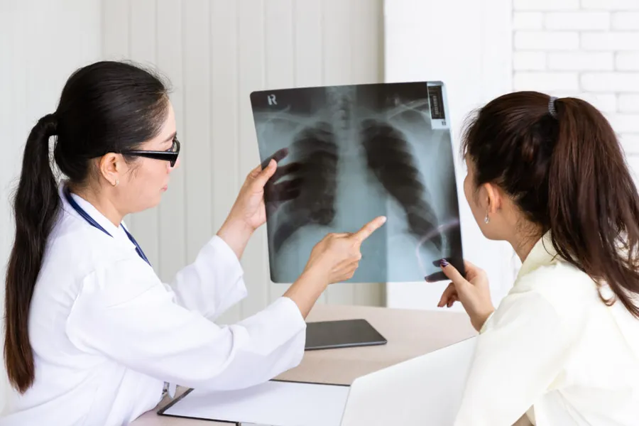
An assessment of the physical structures of the lungs and chest cavity, able to detect early-stage tuberculosis and abnormalities in the lungs, heart, and spine
Benefits of This Test
- Screen for tuberculosis and lung abnormalities at an early stage
- Assess heart size, masses, or foreign objects in the chest cavity
- Detect spinal curvature or old scarring
Testing Procedure
1. Confirm Identity
Provide your information to staff before the X-ray
2. Remove Accessories
Remove jewelry or metal objects from your torso
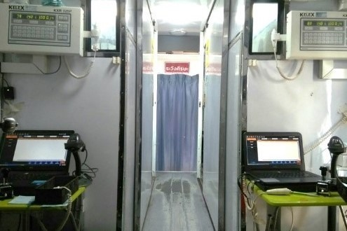
3. X-Ray Capture
Follow staff instructions during the exam
If an abnormality is found
- White patches on the lung:
- You should see a specialist (internal medicine, pulmonology) for further diagnosis
- Enlarged heart:
- Often found in those with high blood pressure or blocked arteries from fat buildup, which makes the heart work harder. If clearly enlarged, see a cardiologist for further diagnosis (note: incomplete inhalation during the X-ray can also make the heart appear larger than it is)
- Curved spine (scoliosis):
- Found at any age, most often in growing teenagers (more common in females) or in adults as the body ages. If mild or painless, seeing a doctor isn't necessary; if severe, an orthopedic specialist is recommended
- Old lesion or calcification:
- A trace of previous internal inflammation that the body has since healed — not a health risk
Tips for the chest X-ray
- Avoid wearing clothing with any metal parts, or an underwire bra for women
- Remove everything from pockets or the chest area to prevent metal from obscuring any findings
Who should not take a chest X-ray
- Those who are pregnant or suspect they may be pregnant should not be X-rayed